
小鼠 单克隆 (SVP-38)
反应物种: 人类, 大鼠
应用: 免疫印迹, 免疫组化
反应物种: 人类, 大鼠
应用: 免疫印迹, 免疫组化


domestic rabbit 多克隆
反应物种: 人类, 小鼠, 大鼠
应用: 免疫印迹
反应物种: 人类, 小鼠, 大鼠
应用: 免疫印迹


Figure 1. Western blot analysis of Synaptophysin using anti- Synaptophysin antibody (PB9409). Electrophoresis was performed on a 5-20% SDS-PAGE gel at 70V (Stacking gel) / 90V (Resolving gel) for 2-3 hours. The sample well of each lane was loaded with 50ug of sample under reducing conditions. Lane 1: rat brain tissue lysates, . Lane 2: mouse brain tissue lysates. After Electrophoresis, proteins were transferred to a Nitrocellulose membrane at 150mA for 50-90 minutes. Blocked the membrane with 5% Non-fat Milk/ TBS for 1.5 hour at RT. The membrane was incubated with rabbit anti- Synaptophysin antigen affinity purified polyclonal antibody (Catalog # PB9409) at 0.5 ug/mL overnight at 4°C, then washed with TBS-0.1%Tween 3 times with 5 minutes each and probed with a goat anti-rabbit IgG-HRP secondary antibody at a dilution of 1:10000 for 1.5 hour at RT. The signal is developed using an Enhanced Chemiluminescent detection (ECL) kit (Catalog # EK1002) with Tanon 5200 system. A specific band was detected for Synaptophysin at approximately 38KD. The expected band size for Synaptophysin is at 34KD.
规格: 100μg/vial
价格: 315美元
至产商
domestic rabbit 单克隆 (BIE-19)
反应物种: 人类, 小鼠, 大鼠
应用: 免疫印迹, 免疫组化, 免疫细胞化学, 免疫沉淀, 流式细胞仪
反应物种: 人类, 小鼠, 大鼠
应用: 免疫印迹, 免疫组化, 免疫细胞化学, 免疫沉淀, 流式细胞仪

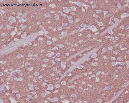
Immunohistochemical analysis of paraffin-embedded human kidney, using Synaptophysin Antibody (M05049). SYP was detected in paraffin-embedded tissue section. Heat mediated antigen retrieval was performed in citrate buffer (pH6, epitope retrieval solution) for 20 mins. The tissue section was blocked with 10% goat serum. The tissue section was then incubated with 1ug/ml rabbit anti-SYP Antibody (M05049)overnight at 4℃. Biotinylated goat anti-rabbit IgG was used as secondary antibody and incubated for 30 minutes at 37℃. The tissue section was developed using Strepavidin-Biotin-Complex (SABC)(Catalog # SA1022) with DAB as the chromogen.

Figure 2. Western blot analysis of Synaptophysin using anti- Synaptophysin antibody (M05049). Electrophoresis was performed on a 5-20% SDS-PAGE gel at 70V (Stacking gel) / 90V (Resolving gel) for 2-3 hours. The sample well of each lane was loaded with 50ug of sample under reducing conditions. Lane 1: rat brain tissue lysates, . Lane 2: mouse brain tissue lysates. After Electrophoresis, proteins were transferred to a Nitrocellulose membrane at 150mA for 50-90 minutes. Blocked the membrane with 5% Non-fat Milk/ TBS for 1.5 hour at RT. The membrane was incubated with rabbit anti- Synaptophysin antigen affinity purified polyclonal antibody (Catalog # M05049) at 0.5 ug/mL overnight at 4°C, then washed with TBS-0.1%Tween 3 times with 5 minutes each and probed with a goat anti-rabbit IgG-HRP secondary antibody at a dilution of 1:10000 for 1.5 hour at RT. The signal is developed using an Enhanced Chemiluminescent detection (ECL) kit (Catalog # EK1002) with Tanon 5200 system. A specific band was detected for Synaptophysin at approximately 38KD. The expected band size for Synaptophysin is at 38KD.
规格: 100微升
价格: 315美元
至产商
domestic rabbit 单克隆 (BGB-19)
反应物种: 人类, 小鼠, 大鼠
应用: 免疫印迹, 免疫组化, 免疫细胞化学
反应物种: 人类, 小鼠, 大鼠
应用: 免疫印迹, 免疫组化, 免疫细胞化学
Immunohistochemical analysis of paraffin-embedded mouse brain, using Synaptophysin Antibody (M05049-1). SYP was detected in paraffin-embedded tissue section. Heat mediated antigen retrieval was performed in citrate buffer (pH6, epitope retrieval solution) for 20 mins. The tissue section was blocked with 10% goat serum. The tissue section was then incubated with 1ug/ml rabbit anti-SYP Antibody (M05049-1)overnight at 4 C. Biotinylated goat anti-rabbit IgG was used as secondary antibody and incubated for 30 minutes at 37 C. The tissue section was developed using Strepavidin-Biotin-Complex (SABC)(Catalog # SA1022) with DAB as the chromogen.

Western blot analysis of Synaptophysin expression in SH-SY5Y cell lysate (M05049-1). Electrophoresis was performed on a 5-20% SDS-PAGE gel at 70V (Stacking gel) / 90V (Resolving gel) for 2-3 hours. The sample well of each lane was loaded with 50ug of sample under reducing conditions. After Electrophoresis, proteins were transferred to a Nitrocellulose membrane at 150mA for 50-90 minutes. Blocked the membrane with 5% Non-fat Milk/ TBS for 1.5 hour at RT. The membrane was incubated with rabbit anti-SYP monoclonal antibody (Catalog # M05049-1) overnight at 4 C, then washed with TBS-0.1%Tween 3 times with 5 minutes each and probed with a goat anti-rabbit IgG-HRP secondary antibody at a dilution of 1:10000 for 1.5 hour at RT. The signal is developed using an Enhanced Chemiluminescent detection (ECL) kit (Catalog # EK1002) with Tanon 5200 system. A specific band was detected for SYP
规格: 100微升
价格: 315美元
至产商
小鼠 单克隆 (3G12)
反应物种: 人类, 小鼠, 大鼠
应用: 免疫印迹
反应物种: 人类, 小鼠, 大鼠
应用: 免疫印迹
微博分享
关注我们的微博
- 来邦网
- 英文来邦