
domestic rabbit 多克隆
反应物种: 小鼠, 大鼠
应用: 免疫印迹, 免疫组化-石蜡切片
反应物种: 小鼠, 大鼠
应用: 免疫印迹, 免疫组化-石蜡切片

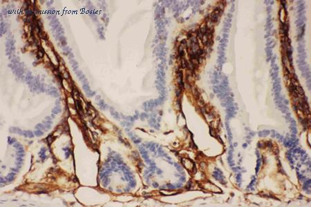
Figure 1. IHC analysis of ICAM1 using anti-ICAM1 antibody (PB9018). ICAM1 was detected in paraffin-embedded section of mouse intestine tissues. Heat mediated antigen retrieval was performed in citrate buffer (pH6, epitope retrieval solution) for 20 mins. The tissue section was blocked with 10% goat serum. The tissue section was then incubated with 1ug/ml rabbit anti-ICAM1 Antibody (PB9018) overnight at 4°C. Biotinylated goat anti-rabbit IgG was used as secondary antibody and incubated for 30 minutes at 37°C. The tissue section was developed using Strepavidin-Biotin-Complex (SABC)(Catalog # SA1022) with DAB as the chromogen.

Figure 2. IHC analysis of ICAM1 using anti-ICAM1 antibody (PB9018). ICAM1 was detected in paraffin-embedded section of mouse lung tissues. Heat mediated antigen retrieval was performed in citrate buffer (pH6, epitope retrieval solution) for 20 mins. The tissue section was blocked with 10% goat serum. The tissue section was then incubated with 1ug/ml rabbit anti-ICAM1 Antibody (PB9018) overnight at 4°C. Biotinylated goat anti-rabbit IgG was used as secondary antibody and incubated for 30 minutes at 37°C. The tissue section was developed using Strepavidin-Biotin-Complex (SABC)(Catalog # SA1022) with DAB as the chromogen.

Figure 3. IHC analysis of ICAM1 using anti-ICAM1 antibody (PB9018). ICAM1 was detected in paraffin-embedded section of mouse spleen tissues. Heat mediated antigen retrieval was performed in citrate buffer (pH6, epitope retrieval solution) for 20 mins. The tissue section was blocked with 10% goat serum. The tissue section was then incubated with 1ug/ml rabbit anti-ICAM1 Antibody (PB9018) overnight at 4°C. Biotinylated goat anti-rabbit IgG was used as secondary antibody and incubated for 30 minutes at 37°C. The tissue section was developed using Strepavidin-Biotin-Complex (SABC)(Catalog # SA1022) with DAB as the chromogen.
规格: 100μg/vial
价格: 315美元
至产商
domestic rabbit 多克隆
反应物种: 人类, 小鼠, 大鼠
应用: 免疫印迹, 酶联免疫吸附测定, 免疫细胞化学, 流式细胞仪, 免疫组化-石蜡切片
反应物种: 人类, 小鼠, 大鼠
应用: 免疫印迹, 酶联免疫吸附测定, 免疫细胞化学, 流式细胞仪, 免疫组化-石蜡切片

Figure 1. Western blot analysis of ICAM1 using anti-ICAM1 antibody (A00171). Electrophoresis was performed on a 5-20% SDS-PAGE gel at 70V (Stacking gel) / 90V (Resolving gel) for 2-3 hours. The sample well of each lane was loaded with 50ug of sample under reducing conditions. Lane 1: rat spleen tissue lysate,. Lane 2: rat thymus tissue lysate,. Lane 3: rat RH35 cell lysate,. Lane 4: mouse spleen tissue lysate,. Lane 5: mouse thymus tissue lysate,. Lane 6: mouse HEPA1-6 cell lysate,. Lane 7: mouse heart tissue lysate. After Electrophoresis, proteins were transferred to a Nitrocellulose membrane at 150mA for 50-90 minutes. Blocked the membrane with 5% Non-fat Milk/ TBS for 1.5 hour at RT. The membrane was incubated with rabbit anti-ICAM1 antigen affinity purified polyclonal antibody (Catalog # A00171) at 0.5 ug/mL overnight at 4°C, then washed with TBS-0.1%Tween 3 times with 5 minutes each and probed with a goat anti-rabbit IgG-HRP secondary antibody at a dilution of 1:10000 for 1.5 hour at RT. The signal is developed using an Enhanced Chemiluminescent detection (ECL) kit (Catalog # EK1002) with Tanon 5200 system. A specific band was detected for ICAM1 at approximately 90KD. The expected band size for ICAM1 is at 58KD.
规格: 100µg/vial
价格: 315美元
至产商
小鼠 单克隆 (LB-2)
反应物种: 大鼠
应用: 流式细胞仪
反应物种: 大鼠
应用: 流式细胞仪
Anti-Human CD54 Icam1 Monoclonal Antibody APC Conjugated, Flow Validated
武汉博士德生物工程有限公司
目录: FC00171-APC
武汉博士德生物工程有限公司
目录: FC00171-APC
小鼠 单克隆 (LB-2)
反应物种: 大鼠
共轭标签: APC
应用: 流式细胞仪
反应物种: 大鼠
共轭标签: APC
应用: 流式细胞仪
小鼠 单克隆 (LB-2)
反应物种: 大鼠
共轭标签: PE
应用: 流式细胞仪
反应物种: 大鼠
共轭标签: PE
应用: 流式细胞仪
微博分享
关注我们的微博
- 来邦网
- 英文来邦